Staff

Richard A. West, M.S.
Specialty: Flow Cytometry, Cell Sorting
Flow Cytometry Core Manager / Associate Scientist
Flow Cytometry Core
Center for Biomedical & Brain Imaging, Room 244
Newark, DE 19716
Lab: 302-831-0627
Cell: 616-795-6928
richwest@udel.edu

Located on the University of Delaware’s main campus in the Center for Biomedical and Brain Imaging (CBBI) and on the STAR campus at AP BioPharma, the UD Flow Cytometry Core Facility offers a full complement of flow cytometry and cell sorting services:
- Immunophenotyping
- DNA Content Analysis
- Veterinary Hematology
- Sample Processing
- Figure Preparation
- Data Analysis
- Consultation
- Assay Design
- Cell Culture
- Cell Sorting
We also offer training, workshops and seminar events that increase skills and knowledge about cytometry and cell biology.
BD FACSAria Fusion High Speed Cell Sorter at CBBI
This state-of-the-art high speed cell sorter features:
- Sterile Biosafety Level II Sorting within HEPA filtered safety cabinet
- 4 laser lines: 488nm Blue, 405nm Violet, 561nm Yellow/Green, 640nm Red
- Detection of up to 15 colors in addition to FSC + SSC
- Sort 4 bulk populations simultaneously or single cell populations into 96/364-well plates
- 98% live cell purity under ideal conditions (viability dye use encouraged)
- 3 configurations: 70um nozzle at 70psi (40 million cells/hour), 85um nozzle at 45psi (25 million cells/hour), or 100um nozzle at 20psi (10-15 million cells/hour)
*Please check with core facility staff to make sure your sample can be sorted on the instrument. There is a nominal half hour charge associated with performing aseptic sorting.*

BD Accuri C6 Plus Flow Cytometer
The Accuri C6 is a walk-up, easy to use, 4-color bench top flow cytometer.
- The system is equipped with a blue laser and a red laser, two scatter detectors and four fluorescence detectors with interference filters optimized for the detection of FITC, PE, PerCP-Cy™5.5 and APC.
- A compact optical design, fixed alignment and pre-optimized detector settings.
- A unique low-pressure pumping system drives the fluidics.
- A sheath-focused core enables event rates of up to 10,000 events per second and a sample concentration over 5 x 10⁶ cells per mL.
- BD Accuri C6 Plus software has an intuitive user interface.
- The tabbed interface provides quick access to the collection, analysis and statistics functions.
- Analysis can be performed on the BD Accuri C6 Plus flow cytometer or can be exported into third-party programs.
- Walk up use is permitted and encouraged.

BD FACSAria Fusion High Speed Cell Sorter (AP BioPharma)
This state-of-the-art high speed cell sorter features:
- Sterile Biosafety Level II Sorting within HEPA-filtered safety cabinet
- 4 laser lines: 488nm Blue, 405nm Violet, 561nm Yellow/Green, 640nm Red
- Detection of up to 15 colors in addition to FSC + SSC
- Sort 4 bulk populations simultaneously or single cell populations into 96/364-well plates
- 98% live cell purity under ideal conditions (viability dye use encouraged)
- 3 configurations: 70um nozzle at 70psi (40 million cells/hour), 85um nozzle at 45psi (25 million cells/hour), or 100um nozzle at 20psi (10-15 million cells/hour)
*Please check with core facility staff to make sure your sample can be sorted on the instrument. There is a nominal half hour charge associated with performing aseptic sorting.*

BD FACSCalibur 4-color Flow Cytometer
The BD FACSCalibur benchtop flow cytometer has the following features:
- 4-color, 6 parameters (Forward and Side Scatter, FITC, PE, PerCP and APC channels)
- Useful for immunophenotyping and DNA content analysis
- CellQuest Pro and ModFit LT Software Applications
- Equipped with low-speed mechanical sort option
- Can analyze up to 5,000 cells per second

VetScan HM5c Hematology Analyzer
The VETSCAN HM5c is a fully-automated, five-part differential hematology analyzer displaying a comprehensive 22-parameter complete blood count (CBC) with cellular histograms on an easy-to-read touch-screen.
Measures WBC (% and absolute count for Lymphs, Monos, Neut, Eos, Baso), RBC (HGB, HCT, MCV, MCH, MCHC), PLT
For use with murine (rodent), canine, equine, bovine, feline (15+ additional species) veterinary blood, marrow, acites and CSF samples (including NHP*)
Can also be used to perform automated counting of cell suspensions from culture or dissociated tissue
*Staff must be informed if you intend to use NHP blood, only approved BSL 2 work is allowed under any circumstances.*

Nexcelom Cellometer Vision CBA Image Cytometry System
All-in-One SystemBasic cell counting, primary cell viability, and cell-based assays.
Dual-Fluorescence for accurate primary cell viability (no interference from red blood cells)
Analyze bone marrow, peripheral blood, and cord blood without lysing
Unique algorithms for advanced cell analysis
Determine concentration and viability of hepatocytes, adipocytes, and other sophisticated cell types
Fast results: Obtain cell images, counts, size measurements, viability calculations, and population data in <3 minutes.


Additional amenities at the Flow Cytometry Core Facility include:
- Common FACS / Flow Cytometry Reagents
- Laminar Flow Hood / BSC Level II
- Sample Prep / Wet Lab
- Refrigerators, Freezer
- Cell Culture Suite
- Centrifuges
- Ice Maker

Dedicated computer workstations allow users to analyze data in the lab. Software packages include:
- FACSDiva 8.0.3
- CFlow Plus / CFlow Sampler
- FCS Express 5.0
- CellQuest Pro 6.0.2
- ModFit LT 2.0
Zeiss Axiovert 200
The Zeiss Axiovert 200 is an inverted epi-fluorescence microscope. Images can be aquired with the AmScope MU-1000 USB 3.0 and AmScope Software or Hamamatsu ORCA-ER digital camera and AxioVision software. The ORCA-ER digital camera is a high resolution gray scale camera.
Standard objectives on this system include:
- 10x Plan Apochromat, 0.45, Ph1
- 20x Plan Apochromat, 0.8, Ph2
- 40x C-Apochromat, 1.2W, oil, DIC
- 63x Plan-Apochromat, 1.4, oil, DIC
Also located in the Multimodal Imaging Suite at CBBI:

Zeiss LSM780 Confocal Microscope
The Zeiss LSM 780 is an upright confocal with 32 channel ultra sensitive GaAsP detectors that allow visualization of weak fluorescent signals with lower photobleaching and phototoxicity. In addition, the system can be used for spectral imaging, photon counting and fluorescence correlation spectroscopy (FCS). The added Multiphoton laser is a 3 Watt Chameleon Vision II with a range of 680 - 1080 nm. The NDD detection system can be used for imaging multiphoton florescence or detection of second harmonic generation (SHG) in the forward and reverse directions. Furthermore, the new OSCiscan function allows faster acquisition of high quality data and better time resolution.
Laser Lines: 405 nm, 458 nm, 488 nm, 514 nm, 561 nm, 633 nm
Upright Microscope, Chameleon Vision II Multiphoton Laser, Reverse and Forward NDD, Fluorescence Correlation Spectroscopy (FCS), Spectral Imaging, Deck Add-On for Intravital Imaging, Photon Counting

Bruker Micro-CT
The SkyScan 1276 is a high performance, stand-alone, fast, desk-top in vivo micro-CT with continuously variable magnification for scanning small laboratory animals (mice, rats, hamster, etc.) and biological samples.
It features a combination of high resolution, big image size, round/spiral scanning, reconstruction, and low dose imaging.
The image field of view (up to 80 mm wide and more than 300 mm long) allows full body mouse and rat scanning.
The variable magnification allows scanning bone and tissue samples with high spatial resolution down to a 2.8 µm pixel size.
Variable X-Ray energy combined with a range of filters ensures optimal image quality for diverse research applications from lung tissue to bone with metal implants.
The SkyScan 1276 in vivo micro-CT administers low radiation dose to the animals allowing multiple scans in longitudinal preclinical studies without the risk of unwanted radiation-induced side effects.
The system can perform scanning with continuous gantry rotation and in step-and-shoot mode with the fastest scanning cycle 3.9 sec.
Attached, dedicated gas anesthesia unit for intravital imaging.

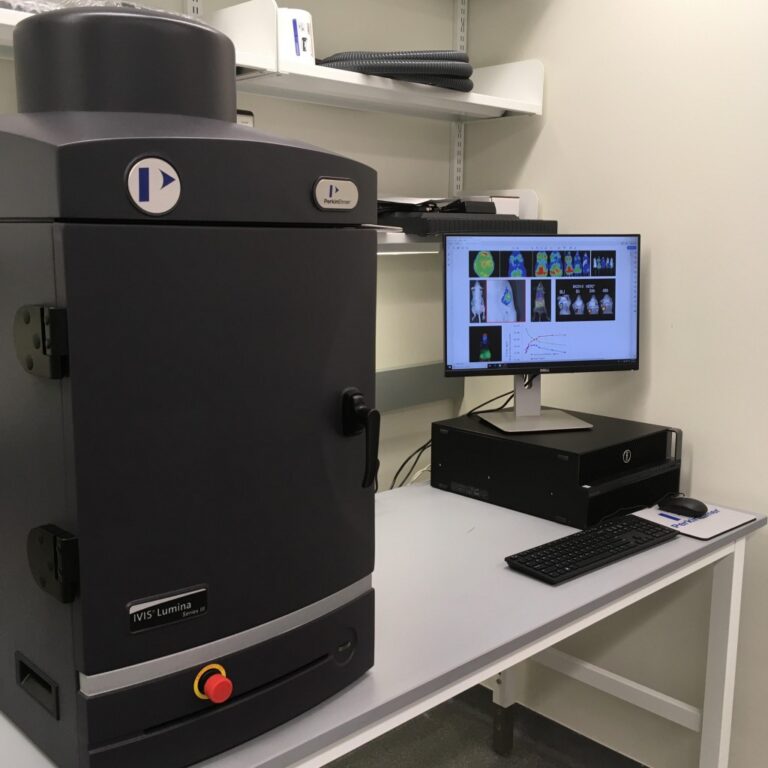
IVIS Lumina III In Vivo Imaging System
The IVIS ® Lumina Series III brings together years of leading optical imaging technologies into one easy to use and exquisitely sensitive bench-top system. The IVIS Lumina III is capable of imaging both fluorescent and bioluminescent reporters. The system is equipped with up to 26 filter sets that can be used to image reporters that emit from green to near-infrared. Superior spectral unmixing can be achieved by Lumina III’s optional high resolution short cut off filters.
Features:
- Exquisite sensitivity in bioluminescence
- Full fluorescence tunability through the NIR spectrum
- Compute Pure Spectrum spectral unmixing for ultimate fluorescence sensitivity
- Dedicated XGI-8 Gas Anesthesia System